Thursday, March 9, 2023

Digestive & Respiratory

So. This week is almost 100% review. There are only TWO totally new structures, and a couple things we've glossed over before, but we're focusing on more this week. Neither of the two new structures (dust cells and Von Kupffer cells) are very easy to find on your own slides, especially the Von Kupffer cells, so I'd for sure suggest finding those with a TA. The material for this week is good preparation for the final, in that we're looking at entire structures and everything we can find in them. Take advantage of this time to study for the final! It's coming up pretty soon and cramming for it will probably kill you.

The Two Totally New Things
There are two macrophages, one in the respiratory system and one in the digestive system, that we haven't learned before.

Von Kupffer Cells

First, in the liver, there are Von Kupffer cells:


A Von Kupffer cell in this slide is labeled "macrophages"

Von Kupffer cells are labeled with arrows in this slide. Note also the binucleated cells, which we'll only see in the liver

For Von Kupffer cells, what we're looking for are cells that line the sinusoids of the liver, and whose nuclei protrude out into the sinusoid. Sometimes their nuclei are darker and smaller, like the second slide, but more often they are larger than the other hepatocyte nuclei. It just depends on the slide. They are MODIFIED MONOCYTES histologically. (Or modified simple squamous, but I think modified monocytes is easier to remember because they're macrophages specialized to liver tissue.)

Dust Cells
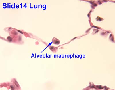
The second ones are called dust cells, and we find those guys in the lungs:

A dust cell is labeled as an "alveolar macrophage" on this slide

Dust cells are labeled in these slides with the black arrows

For dust cells, we're looking for cells with large nuclei, on their own, close to the border of the alveolus. For uniformity in grading the u-finds, the TA's are looking for cells that don't touch the simple squamous epithelium of the alveoli, but are found close to it. Sometimes little simple squamous cells will be separated from the alveolar border, and students will point to those thinking they're dust cells, but they're not. So make sure the cell you're pointing to a cell with a nice round, big nucleus (big compared to the simple squamous cells). They are also MODIFIED MONOCYTES histologically.


The Kind-of-New Things
We have talked about the mucosal layers before, but we haven't really focused on them. Make sure you know them for this upcoming quiz.

Mucosal layers are shown by brackets. This is the duodenum -- note the Brunners glands in the submucosa
A: Mucosa                  
B: Muscularis Mucosa
C: Submucosa            
D: Muscularis externa 

Obviously this slide shows the layers perfectly, but it won't always be the case in your own slides. The small intestine is the easiest area to see all these layers, but keep in mind they are found throughout the GI and respiratory tracts. The muscularis mucosa layers separate the mucosa from submucosa, and the muscularis externa is the one that includes the muscle layers (inner circular, outer longitudinal, for example).


Going through the digestive system, there are a few things that are kind of new-ish.

You'll notice that this week we taught you guys the muscle layers surrounding the esophagus. It's pretty intuitive though since it is part of the GI tract: Inner circular, outer longitudinal.
Always keep in mind the relative anatomy of the structures we're learning about! Which is the trachea? Which is the esophagus? What would it look like if a pic of the esophagus included part of the trachea?

Make sure you know how to distinguish between the different parts of the small intestine: duodenum (Brunner's Gland), Jejunum (nothing remarkable), Ileum (Peyer's Patches) [pictures below in that same order]


Gallbladder - We've never talked about this guy before, but don't worry too much about it. We're pointing it out here because it's part of the digestive system, but it won't ever be a u-find or anything. It has evaginations and the simple columnar epithelium, but it doesn't have the striated border like the small intestine. Pretty unique looking. Because it's right up next to the liver, you can often see the liver in the slide as well, like in this slide:


The gallbladder here is the main structure that looks like Lake Powell kind of. The dense purple tissue around the edges of the slides, is the liver


Keep in mind this week that everything you've learned within the digestive and respiratory tracts are all fair game. Remembering things like trends for goblet cell concentration (increases down the digestive tract and decreases down the respiratory tract) will help you recognize the structures you're in. If I were you, I would google things like "ileum histology" and name everything you can on the slides that come up.

For example:
Ileum

-Simple columnar epithelium with a striated border
-Goblet cells, which secrete mucin proteoglycans and are unicellular exocrine glands histologically
-Paneth cells, which secrete antimicrobial enzymes, are found in the crypts of the evaginations, and    are also unicellulare exocrine glands histologically
-Lamina propria, which is loose connective tissue histologically, are found inside the evaginations
-Mucosa is the layer that contains all those things
-Muscularis mucosa separates mucosa from submucosa
-Submucosa, mostly made up of loose connective tissue and here contains peyer's patches
-Peyer's patches, which are made up of dense lymphatic tissue histologically and contain germinal  centers, and are how we identify this slide as the ileum specifically
-Muscularis externa, which here includes the inner circular and outer longitudinal muscle layers,  which are both smooth muscle regularly arranged histologically
-Aurebach's and Meissner's plexus could both theoretically be found in this slide, though we usually  look for them in the jejunum. Their function is to regulate peristalsis. 


Stuff from this section that won't be u-finds:
-G cells
-Gallbladder
-Larynx
Stuff from this section that you won't need to know for this week (but will in the future):
N/A
Stuff from this section you don't need to know for lab:
-Pages 55 and 59 are helpful to know, but you won't be quizzed on them directly
-Larynx. Don't worry about it

Thursday, March 2, 2023

Endocrine Stuff

Friends, with the conversion of our lab to online please use all resources including this blog. TA's will have office hours through zoom (details forthcoming), use the  PDBio325 website for practice quizzes, slide databases, powerpoints, and other review material. Remember, since we do not have U-Finds and We-Finds expect more slide questions.

There is quite a bit of memorization for this week. Good luck. The good thing is learning this stuff really well will help you in almost every bio class you have here on out. This week the anatomical structures aren't too difficult, but make sure you know what hormone(s) they all secrete, the effects of those hormones and how they all interact with each other (i.e. what stimulates/works with/inhibits what). Really get to know the table on page 54 of your lab book.


Pituitary Gland

A little something about the pituitary gland: 
Note: this purple text stuff is more detail than you need to know for the lab, but understanding this helps make sense of the different pars and why they're named what they are. 
During development, the posterior pituitary (henceforth referred to as the neurohypophysis) grows down from the brain. The anterior pituitary (adenohypophysis) grows up from the roof of the mouth (that connection later degenerates). So the neurohypophysis is nerve tissue, but the adenohypophysis is like its own little organ that's close enough to they hypothalamus to receive orders from it. This helps when you're trying to figure out the different pars. The neurohyphophysis is made up of pars nervosa (which makes sense because it's nervous tissue). The adenohypophysis is made up of pars distalis (you can think of it growing up from a different structure, so the tissue in there is distantly related to pars nervosa). 


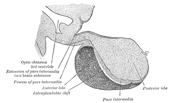
Illustration of pituitary gland, showing the different pars


As the adenohypophysis grows up to meet the neurohypophysis, the capsule around the adenohypophysis reaches the infundibulum and wraps around it, to anchor the adenohypophysis to that spot. So you can see in the picture above that the pars intermedia and the pars tuberalis are really the same tissue, just in different spots. In fact, some texts just call the pars tuberalis the "process of pars intermedia." 

Image result for pituitary gland pars

What we would normally call "pars tuberalis" is referred to in this text as "process of pars intermedia" and "extension of pars intermedia into brain stubstance"


All you really need to know about the different pars for the lab is that the adenohypophysis is pars distalis and the neurohypophysis is pars nervosa. 

Adenohypophysis:
Choromophobic chromophils don't have an acronym, because they don't make any hormones
B-FLAT - Basophilic chromophils secrete FSH, LSH, ACTH, and TSH
GAP - Acidophilic chromophils secrete GH and Prolactin

Make sure you know ALL of the hormones that are secreted by each of the cells in the different organs and what each of those hormones does.

Also, definitely know how to identify each of the 3 cell types found in the pars distalis ON YOUR OWN SLIDES. They look really clear in your lab manual, but it may or may not look like that on your slide.

Keep in mind while looking at your own pituitary slides that depending on where yours is cut, it may or may not have the infudibulum/pars tuberalis on there. It may also have mostly adenohypophysis with only a little neurohypophysis, or the other way around. Make sure you're oriented in your own slide.


All three pictures here show the division between the adenohypophysis and neurohypophysis, What cells can you see in the last picture? You should be able to point out FIVE kinds and know what they secrete. 

See the 3 different types of cells? Which ones are the acidophilic chromophils? Basophilic? Remember, cells that are acidophilic tend to stain brightly, or appear more 'warm' in color

Neurohypophysis: 
While the adenohypophysis does a lot of synthesizing and releasing of many different hormones, all the neurohypophysis does is store and release the oxytocin and antidiuretic hormone (ADH, also known as vasopressin and AVP in some texts) produced in the hypothalamus. 

If you look at the structures of these two molecules, you'll notice they're very similar (not that you'll ever have to known the structures in this class). This visual helps me remember that these guys both come from the neurohypophysis. 

These two hormones are gathered and stored in what's called "herring bodies," whose appearance varies a LOT depending on the slide. 

In this one, the herring bodies are areas that look slightly pinker than the cytoplasm of the pituicytes. Note the lack of nuclei within the herring bodies

If your slide looks like this, you're lucky. These herring bodies are very dark compared to everything else. They're all those red spots

The herring bodies in this one are pretty much the same color as the pituicytes. The biggest tipoff for herring bodies in this one is just areas that lack nuclei


Thyroid/Parathyroid

Just a little anatomy for you:

So now if we zoom in on the thyroid gland, we see this circle-y organization, with the circles filled in with stuff. One thing the thyroid can be easily confused with are lactating mammary glands, but the thyroid gland will have cuboidal cells around the colloid matter, and their nuclei will be round and defined. In lactating mammary glands, there will be a lot more connective tissue, which, in the context of a lactating gland, means you won't really see nice circular defined nuclei like this. 

Thyroid hisology

In the slide above, we can see both follicular cells and parafollicular cells. Follicular cells will be any of those touching the colloid matter (the purple stuff with no nuclei in the above slide), and parafollicular cells will be those in between. 

Keep in mind that follicular cells DO NOT secrete T3 and T4. They secrete thyroglobulins, which are later converted to t3 and t4 in the colloid matter by enzymes found in there. 

Because the parathyroid lays right on the thyroid, you can definitely have slides with both structures on them:


Thyroid (upper stuff) and parathyroid (lower stuff)

 Thyroid and parathyroid (dark purple)


Thyroid and parathyroid

Thyroid (A) and parathyroid (B)
Zooming in on the parathyroid, we can distinguish two kinds of cells: principal cells, whose nuclei will stain lighter, and oxyphil cells, whose nuclei are typically a little smaller and darker/denser.

Parathyroid - You can see principal/chief cells, oxyphil cells and unilocular adipocytes in this shot. Can you point each of them out? Remember that principal cells are the smartest, look like brains (chromatin) and the oxyphil cells are more dense looking.


Pancreas

Islet of Langerhans in the pancreas

Your book is pretty clear on these. Make sure you know where to find alpha, beta, delta, and PP cells and what they all secrete. Delta cells inhibit the endocrine function of the pancreas while PP cells inhibit the exocrine function. 


Adrenal Gland
Know the layers of the adrenal gland! It's easy to identify the medulla, then if asked to point to a specific layer (zona glomerulosa, zona fasciculata, zona reticularis), point to the outside, middle or inside of the cortex, respectively. Know what each zone secretes (both the general class of hormones and a specific example, e.g. glucocorticoids and cortisol). The four pictures that follow are all of the same gland.

Human adrenal gland, way zoomed out

Zoomed in... Can you name the three layers of the cortex and find the medulla?


See the medulla in here?
Trick question! There is no medulla in this shot, just doubled up cortex. See the pic below for an explanation.


It is a very good idea to spend some quality time with the adrenal gland -- not only is there a deceptively large amount of memorization associated with it, but it will reappear in almost every bio class from here on out, for reals. 

Some mnemonics: 
Great Men Always (glomerulosa, mineralocorticoids, aldosterone)
Find Cute Girls (fasciculata, cortisol, glucocorticoids) 
Really Darn Attractive (reticularis, DHEA, androgens)
This one's not the most wonderful, because they're in different orders, you know what I mean?

Great Men Always (glomerulosa, mineralocorticoids, aldosterone)
Find Great Cowboys (fasciculata, glucocorticoids, cortisol)
Riding A Donkey (reticularis, androgens, DHEA)

Aldosterone controls salt retention, corisol increases blood glucose levels, and DHEA is a precursor to sex hormones like estrogen and testosterone. So from the outside to inside, the layers are in charge of salt, sugar, sex (it gets sweeter as you get deeper ;) )


Argentaffin Cells

You'll find these guys in cross sections of villi in the small intestine. Typically they are darker and wedge shaped (most of your slides will look just like the one in the book), but sometimes a different stain can yield something that looks a little more like this:

In this one, the purple arrow is pointing at an argentaffin cell. Just to show you they don't always look like the one in your book 

Stuff from this section that won't be u-finds:
-Infundibulum -Pars tuberalis
- G-cells (just don't even worry about them at all, in fact)
Stuff from this section that you won't need to know for this week (but will in the future):
N/A
Stuff from this section you don't need to know for lab:
-Page 51 is helpful to know, but you won't be quizzed on it directly


One more time, for the love of all that is good, MAKE SURE you know what everything secretes and what those secretions do. Know the heck out of the table on page 54 of your lab book.


Endocrine Review Sheet
Key: Know the anatomical and histological names (including modifications) for the following bolded structures; assume that you will be required to find the structures indicated by * on your own slides.
***This list is not guaranteed to be exhaustive, and only includes terms from this unit. While we will not focus on quiz information from previous weeks, knowledge of previous material may be useful***
Focus on endocrine histology, but keep in mind all of the following terms. Know the endocrine secretions and functions as outlined in your manual.
Pituitary Gland
Anterior Pituitary (Adenohypophysis)
·       Pars distalis*
·       Chromophobes*
·       Basophilic chromophils*
·       Acidophilic chromophils*
·       Pars intermedia*
·       Pars tuberalis*
Posterior Pituitary (Neurohypophysis)
·       Herring bodies*
·       Pituicytes*


Thyroid
·       Follicular cells*
·       Parafollicular cells*
Parathyroid
·       Principal (chief) cells*
·       Oxyphil cells*
Pancreas
Know the 4 types of cells found in Islets of Langerhans, the general region where they are found, as well as their secretions
·       α-cells
·       β-cells
·       δ-cells
·       PP-cells
Stomach
·       G-cells
·       Parietal cell*
·       Chief cell*
Adrenal Gland
For each region of the adrenal gland, know the class of hormone that gets secreted as well and  a specific hormone example.
·       Zona glomerulosa*
·       Zona fasciculate*
·       Zona reticularis*
·       Medulla*
Enteroendocrine Cells
·       Argentaffin cells*

Thursday, February 16, 2023

Muscle Stuff

Congrats on surviving the midterm (hopefully...) Hopefully the midterm was able to help you see areas you need to practice or helped boost your confidence in what you already know. If you didn't do so hot, remember you can drop the midterm and that there's still the final which is worth half of your grade.

This week is muscle - make sure you know the differences between the 3 types of muscle:

See the difference between the transverse and longitudinal cuts?


Skeletal:
- multinucleated
- peripheral nuclei
- striations
- can be regularly or irregularly arranged


See the layers?


Smooth:
- uninucleated
- central nuclei
- no striations
- fusiform shape
- usually comes in layers


The intercalated discs allow current to pass from cell to cell


Cardiac:
- bifurcated
- central nuclei
- intercalated discs containing several gap junctions
- striations


Also, be sure you can find/identify intrafusal fibers and know their function, i.e. prevention of muscle over-stretching; remember their histological name is modified skeletal muscle fiber and that the capsule is DCCTRA. Normally, intrafusal fibers are about 2-3 times the size of a single skeletal muscle fiber and are surrounded by a collagen sheath (connective tissue).


Sarcomeres

So, just a refresher from anatomy of what's actually going on in the sarcomere - remember that all the actin is coming from the Z-line and all the myosin is coming from the M-line. Contraction is due to an increase in overlap between actin and myosin. The M-line is located in the A-band. The A band doesn't change size during contraction. Remember that it is the I-band and H-bands that shrink as myosin and actin overlap more.


 Now, try to find the above structures on a real image. Remember: 'Zoro Is A Hairy Man'


As we zoom out, can you see where those striations come from? Smooth muscle doesn't look like this because they don't have sarcomeres like skeletal and cardiac muscle; they have a completely different method of contraction which you will learn more about in advanced physiology


 Also, make sure to study exceptions/anomalies - those tend to make good quiz/final questions :) For instance, the tongue is the only place in the body where we find skeletal muscle irregularly arranged.

See the longitudinal AND transverse muscle fibers?


Also, when it comes to memorizing the different smooth muscle layers, remember most of the time the layers will be inner circular, outer longitudinal - then just memorize the exceptions, of which there are a few. Also, remember that the inner circular layer is responsible for segmentation while the outer longitudinal layer is required for peristalsis.

The exceptions are:
1. the stomach - has three layers; essentially just adds an inner oblique layer to the normal inner circular, outer longitudinal - so it becomes inner oblique, middle circular, outer longitudinal
2. the uterus and vagina - girls are all over the place, right?  Almost all of the female female repro system is smooth muscle irregularly arranged - think about the parts of the female repro that have to do a lot of stretching - those are the ones that are going to be irregular
3. the vas deferens - remember that the vas deferens has tons of muscle - it needs that muscle to act as a pump - I think of the vas deferens as a sandwich (inner longitudinal, middle circular, outer longitudinal)
4. the ureter: the ureter is just whacked - it's exactly opposite of everybody else: inner longitudinal, outer circular


Blood Vessels

Make sure you know the anatomical layers of the heart - epicardium, myocardium, endocardium, and what they are histologically

Purkinje fibers - these are modified cardiac myocytes located just under the endocardium in a region called the subendocardium; remember that impulses in the heart originate at the SA node, travel to the AV node, through the left and right bundles located in the interventricular septum to the purkinje fibers - these fibers travel through the ventricular walls to spread the current


- purkinje fibers are non-contractile cells and contain fewer myofibrils - this means they take up less stain, i.e. they are generally lighter on a slide and are larger, often binucleated cells.

Look for them near the 'edge' of the tissue on your slide - if you point to cells in the middle of the myocardium, chances are they're not purkinje fibers - as you can see in the diagram on the left, purkinje fibers tend to stay out of the muscle wall


Large, medium, small artery and capillary


Make sure you know what makes up the different tunics:
1. tunica intima: endothelium + basement membrane + internal elastic lamina
2. tunica media - just the muscle layer
3. tunica adventitia - LCT with some DCCTRA


Large artery:
See the 1:1 muscle cell layer to elastic fiber ratio?

Medium artery:

Notice the distinct internal and external elastic laminas; also, the virtual absence of elastic fibers in the tunica media


Small artery:
Note the absence of an external elastic lamina, but still a distinct internal elastic lamina


Also, know capillaries and where to find them (lungs and kidneys are a good place) and that they are made of simple squamous epithelium.


Large, medium, small veins


 This pic has it all -- notice the differences between the medium artery and medium vein? The vein is much more collapsed and has a much thinner muscle layer - remember, they don't do any pumping, whereas the arteries do lots of pumping and stretching.

Also, veins tend to have a much thicker tunica adventitia and no internal/external elastic laminas

For the purposes of this class, a large vein is one whose lumen doesn't fit entirely on the view screen.

Stuff from this section that won't be u-finds:
-Intrafusal fibers

-Sarcomere Stuff from this section that you won't need to know for this week (but will in the future):
N/A
Stuff from this section you don't need to know for lab:
-Pages 34 and 40 are helpful to know but you will not be quizzed on them directly

-Arterioles
-Venules


Muscle Review Sheet
Key: Know the anatomical and histological names (including modifications) for the following bolded structures; assume that you will be required to find the structures indicated by * on your own slides.
***This list is not guaranteed to be exhaustive, and only includes terms from this unit. While we will not focus on quiz information from previous weeks, knowledge of previous material may be useful***


Focus on muscle histology, but keep in mind all of the following terms.
Skeletal Muscle
Transverse Cut
·       Skeletal muscle*
·       Epimysium*
·       Perimysium*
·       Endomysium*
·       Fascicle*
·       Peripherally located nuclei
Longitudinal Cut
·       Perimysium*
·       Endomysium*


Intrafusal Fiber
·       Intrafusal fibers*
·       Capsule
Sarcomere
·       A-band
·       H-disk
·       M-line
·       Z-line
·       I-band
Neuromuscular Junction
·       Terminal bouton (motor end plate)
·       Nerve axons
·       Skeletal myofibers
Tongue
·       Circumvallate papillae*
·       Von Ebner’s gland
·       Taste bud
·       Skeletal muscle*
Cardiac Muscle
Cardiac Muscle
·       Bifurcations
·       Intercalated discs
·       Centrally located nuclei

Smooth Muscle
Longitudinal Cut
·       Smooth muscle fiber (fusiform shape)
·       Centrally located nuclei

Transverse Cut 
·       Smooth muscle myocyte
·       Centrally located nuclei
 Vasculature 
·       Tunica intima*
·       Tunica media*
·       Tunica adventitia*
Intestine
·       Muscularis externa*
·       Inner circular*
·       Outer longitudinal*
·       Muscularis mucosa*
Stomach
·       Muscularis externa*
·       Inner oblique*
·       Middle circular*
·       Outer longitudinal*
·       Gastric gland
·       Muscularis mucosa*


Reproductive Tract
Uterus
·       Endometrium*
·       Myometrium*
·       Uterine glands
·       Uterine epithelium
Vagina
·       Smooth muscle*
·       Lamina propria
·       Vaginal epithelium
Oviduct
·       Oviduct epithelium
·       Lamina propria
·       Muscularis externa*
·       Inner circular*
·       Outer longitudinal*
Vas Deferens
·       Vas deferens epithelium
·       Inner longitudinal layer*
·       Middle circular layer*
·       Outer longitudinal layer*
Urinary Tract
Ureter
·       Ureter epithelium
·       Inner longitudinal layer*
.         Outer circular layer*

Circulatory System
Heart
Outer Heart Wall
·       Adipose tissue
·       Myocardium*

Inner Heart Wall
·       Endocardium*
·       Purkinje fibers*
Vasculature
Large Artery
·       Tunica intima*
·       Internal elastic lamina*
·       Tunica media*
·       Tunica adventitia*
Medium Artery
·       Tunica intima*
·       Internal elastic lamina*
·       Tunica media* (sporadic elastic fibers)
·       Tunica adventitia*
Small Artery
·       Tunica intima*
·       Tunica media* (~4 layers)
·       Tunica adventitia*
Arteriole
·       Tunica intima* (very thin internal elastic lamina)
·       Tunica media* (~2-3 layers)
·       Tunica adventitia
Capillary
·       Endothelium*
Large Vein
·       Tunica intima (no elastic lamina)*
·       Tunica media*
·       Tunica adventitia*
Medium Vein
·       Tunica intima*
·       Tunica media* (a few muscle layers)
·       Tunica adventitia*