The Two Totally New Things
There are two macrophages, one in the respiratory system and one in the digestive system, that we haven't learned before.
Von Kupffer Cells
First, in the liver, there are Von Kupffer cells:

A Von Kupffer cell in this slide is labeled "macrophages"

Von Kupffer cells are labeled with arrows in this slide. Note also the binucleated cells, which we'll only see in the liver
For Von Kupffer cells, what we're looking for are cells that line the sinusoids of the liver, and whose nuclei protrude out into the sinusoid. Sometimes their nuclei are darker and smaller, like the second slide, but more often they are larger than the other hepatocyte nuclei. It just depends on the slide. They are MODIFIED MONOCYTES histologically. (Or modified simple squamous, but I think modified monocytes is easier to remember because they're macrophages specialized to liver tissue.)
Dust Cells
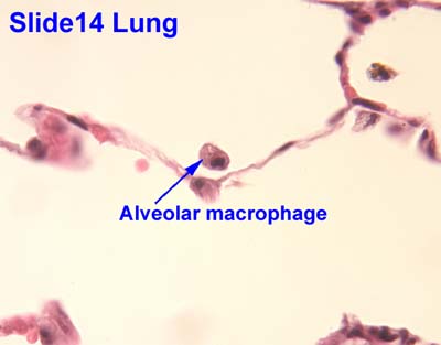 |
| A dust cell is labeled as an "alveolar macrophage" on this slide |

Dust cells are labeled in these slides with the black arrows
The Kind-of-New Things
We have talked about the mucosal layers before, but we haven't really focused on them. Make sure you know them for this upcoming quiz.

Mucosal layers are shown by brackets. This is the duodenum -- note the Brunners glands in the submucosa
A: Mucosa
B: Muscularis Mucosa
C: Submucosa
D: Muscularis externa
B: Muscularis Mucosa
C: Submucosa
D: Muscularis externa
Obviously this slide shows the layers perfectly, but it won't always be the case in your own slides. The small intestine is the easiest area to see all these layers, but keep in mind they are found throughout the GI and respiratory tracts. The muscularis mucosa layers separate the mucosa from submucosa, and the muscularis externa is the one that includes the muscle layers (inner circular, outer longitudinal, for example).
Going through the digestive system, there are a few things that are kind of new-ish.
You'll notice that this week we taught you guys the muscle layers surrounding the esophagus. It's pretty intuitive though since it is part of the GI tract: Inner circular, outer longitudinal.
 |
| Always keep in mind the relative anatomy of the structures we're learning about! Which is the trachea? Which is the esophagus? What would it look like if a pic of the esophagus included part of the trachea? |
Make sure you know how to distinguish between the different parts of the small intestine: duodenum (Brunner's Gland), Jejunum (nothing remarkable), Ileum (Peyer's Patches) [pictures below in that same order]


Gallbladder - We've never talked about this guy before, but don't worry too much about it. We're pointing it out here because it's part of the digestive system, but it won't ever be a u-find or anything. It has evaginations and the simple columnar epithelium, but it doesn't have the striated border like the small intestine. Pretty unique looking. Because it's right up next to the liver, you can often see the liver in the slide as well, like in this slide:

The gallbladder here is the main structure that looks like Lake Powell kind of. The dense purple tissue around the edges of the slides, is the liver
For example:

Ileum
-Simple columnar epithelium with a striated border
-Goblet cells, which secrete mucin proteoglycans and are unicellular exocrine glands histologically
-Paneth cells, which secrete antimicrobial enzymes, are found in the crypts of the evaginations, and are also unicellulare exocrine glands histologically
-Lamina propria, which is loose connective tissue histologically, are found inside the evaginations
-Mucosa is the layer that contains all those things
-Muscularis mucosa separates mucosa from submucosa
-Submucosa, mostly made up of loose connective tissue and here contains peyer's patches
-Peyer's patches, which are made up of dense lymphatic tissue histologically and contain germinal centers, and are how we identify this slide as the ileum specifically
-Muscularis externa, which here includes the inner circular and outer longitudinal muscle layers, which are both smooth muscle regularly arranged histologically
-Aurebach's and Meissner's plexus could both theoretically be found in this slide, though we usually look for them in the jejunum. Their function is to regulate peristalsis.
Stuff from this section that won't be u-finds:
-G cells
-Gallbladder
-Larynx
Stuff from this section that you won't need to know for this week (but will in the future):
N/A
Stuff from this section you don't need to know for lab:
-Pages 55 and 59 are helpful to know, but you won't be quizzed on them directly
N/A
Stuff from this section you don't need to know for lab:
-Pages 55 and 59 are helpful to know, but you won't be quizzed on them directly
-Larynx. Don't worry about it

No comments:
Post a Comment