Not going to lie, this week is pretty heavy on material too. Really work at mastering each week's material, because it will help you not only for that week's quiz, but also for each one that follows. The load lightens up quite a bit here soon, I promise.
Tips for this week:
1. Make SURE you know each gland anatomically and histologically, as well as what organ it's found in (e.g. Brunner's gland (anatomical name) is CMBT (histological name) and is part of the duodenum (small intestine)). Also, be sure to study what each gland secretes. All of these are fair game for the quiz. There is quite a bit of memorization involved with exocrine glands because there isn't a real clear connection between the gland classification (e.g. SMBT) and its function (besides that more active glands are usually alveolar as opposed to tubular).
2. Use the mnemonics we gave you in class to remember the histological names of the glands or, if you are creative and want to make up your own mnemonics, that's fantastic! Feel free to share them and I'll post them here, along with these:
CMTA ('Come meet the acid'): subesophageal, submandibular, and proliferating mammary
UNICELLULAR = paneth and goblet cells
CMBT ('combat') in the Brunner's - think an acid/base 'combat' ;)
SMBT ('smell my big/baby tummy'): smell = olfactory (Bowman's) my big = pregnant (uterine) tummy = (gastric)
CMA ('Country Music Awards') -- we've got Dolly Parton singing with her tongue (von ebner's) into her pancreas microphone, with her large bosoms (lactating mammary) and parotid gland earrings, and with everyone in the audience drinking their beers that accompany country music while their livers cringe.
SMTA ('some men talk a lot') in the sub-tracheal
CMT ('cow milk trapping') = resting mammary
CMBTA ('come men be tested annually') in the prostate
SMCT ('some men can't tell') in the sweat gland
SMBA ('Simba is a dirty lion') remember, the sebaceous gland secretes sebum, which when Rafiki holds Simba up, you can see his bum. Rafiki also spreads that janky oil across Simbas face.
Blue=Histological
Red=Anatomical
Broken up so that each line only contains one histological name with the corresponding anatomical names.
3. Don't memorize things in order! If you only know the material in the order it's presented in the manual, the quizzes will be much harder for you. Try memorizing things by histology - pick a type of gland (say, CMA) and name every gland with that histology (von ebner's, pancreas, parotid, lactating mammary, liver).
4. Finally, I want to emphasize that it is still possible to memorize the ENTIRE lab manual and still fail this class. You gotta spend time practicing identifying structures on slides. Look up images from different organs that you struggle with or that seem identical to you, put them side by side and practice noticing their unique landmarks. Each and every organ has at least one landmark that makes it unique and you can be sure that we will test you on it.
Other things to remember from glands:
-Goblet cells decrease down the respiratory tract and increase down the GI tract --> you would be most likely to find them in the large intestine and trachea and least likely in the esophagus and terminal alveoli. If you're asked to find one on a U-Find, go straight for the large intestine. There are TONS there.
-Sub-esophageal and sub-tracheal glands can look pretty similar, but are obviously distinguished by the epithelium (strat. squamous for sub-esoph. and ciliated pseudo. colum. for sub-trach.) and the presence/lack of cartilage c-rings (sub-trach has the c-rings).
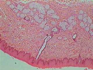

Can you see the difference in the epithelium in the above pics? (Left is esophagus, epithelium on the bottom, right is trachea, epithelium on the top)
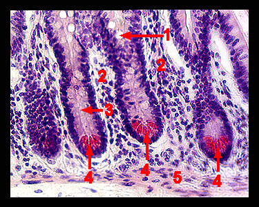
- Paneth cells: DON'T EXPECT TO BE ABLE TO ACTUALLY SEE PANETH CELLS ON YOUR OWN SLIDES. Students get confused about this every semester -- know where they are, but know you won't be able to actually see them. Unless your slide was stained for them. Which it won't be. If you need to point out paneth cells, just point to the intestinal crypts of the small intestine. (Paneth cells are found in the crypts mostly of the small intestines, but may be found sporadically throughout the cecum and appendix. Paneth cells are acidophilic, meaning that IF they are stained for, they typically stain brighter colors (usually red). Also, just something interesting, Paneth cells are located adjacent to stem cells at the base of the intestinal crypts which are responsible for the long-term maintenance of the epithelium. Paneth cells play a role in protecting these stem cells by secreting antimicrobial enzymes.)
 |
-Brunner's gland is only in the duodenum section of the small intestine.
If asked to identify the location of the gland, be as specific as
possible. Don't just say 'small intestine,' put 'duodenum.'
|
-The submandibular/submaxillary glands can sometimes be tricky for people. Look for serous AND mucous acini. The biggest landmark for these are the demilunes (look like a half-moon because of one side of serous and one side of mucous acini)

Submandibular Gland with demilunes
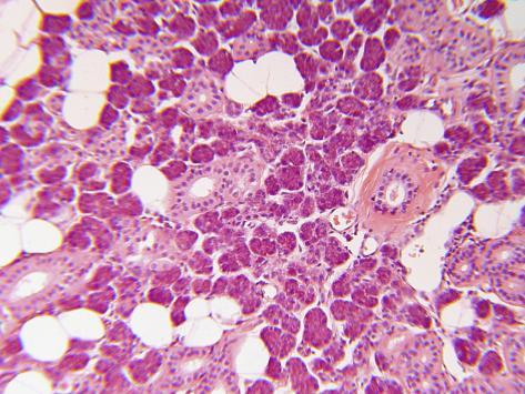 |
| Parotid Gland. White lobules are the adipose. |
 |
| Parotid - see the ducts? |
 |
| Pancreas w/islet of Langerhans |
- Notice how both pictures have areas that are lighter than the surounding acini; however, in the top picture, the lighter cells are obviously cuboidal with a central lumen - those are the ducts of the parotid exocrine gland. The bottom picture on the other hand, has an islet of Langerhans with very non-cuboidal cells arranged in kind of a jumbled, messy way. You can't see any ducts/lumens because this part of the pancreas is endocrine.
- Liver: Although we focused mainly on the hepatic triad when discussing the liver this week, you also need to be able to recognize the liver without the hepatic triad to guide you. The biggest landmark for the liver (besides the triad) are the binucleated hepatocytes. We will always show you at least one binucleated cell. Also, the liver is often split into lobes without any other major structures (besides the hepatic triad).
 |
| Note the binucleated cell in the upper left hand quadrant. Also, can you see the hepatic sinusoids? |
 |
| No triad? Find binucleated cells! |
 |
| There's the triad! Now which structure is which? Remember how you can tell the difference? |
-The biggest landmark for the Bowman's is the epithelium (ciliated pseudo. colum.). It usually is very thick, has an especially thick band of nuclei, and you usually won't see any nuclei at the apical 2/3's.

Nasal epithelium and Bowmans Glands
-For the Uterine gland, remember the analogy of the wild leopard spots :) The uterine gland is truly unique and should be easy points on the quiz if we show you a picture.
-Remember that the mammary glands secrete COLOSTRUM, not milk. If there are lots of follicles and they all look full with colostrum, it is lactating. If there aren't many at all, and they aren't really filled with colostrum, it is resting. In between those two is proliferating (look for about 50/50 filled/empty follicles).
 |
| Resting mammary - what is this histologically??? |
 |
| Lactating Mammary |
-Both the sweat and sebaceous glands are found in the dermis (deep to the epidermis). Remember that the sweat glands are coiled tubular and will have pretty distinct lumens (look like sliced tree trunks) whereas the sebaceous ducts aren't as distinguishable (looks more like a cluster of grapes). The sebaceous glands are oftentimes right by a hair follicle (where they secrete the sebum into).
 |
| Sweat or sebaceous? (starts w/ S and ends in weat...) |
Hope this helps! Remember, if you have any questions or want me to post more pics/address any questions, email me at oxleybk@gmail.com and I will add my responses to the blog. Also, GO TO OPEN LAB at least once this week - it will make all the difference! Good luck!
Stuff from this section that won't be u-finds: - Neck cells (stomach) - Circumvallate papillae - Taste buds
- Interlobular septa (liver) Stuff from this section that you won't need to know for this week (but will in the future): - Muscles referred to in this section (skeletal muscle in tongue, myometrium, etc.) are there for reference, but won't be tested on this week - Hyaline cartilage of trachea - Seminal vesicle
Stuff from this section you don't need to know for lab:
- The whole page 16 is helpful to know, but you won't be quizzed on it directly
Stuff from this section that won't be u-finds: - Neck cells (stomach) - Circumvallate papillae - Taste buds
- Interlobular septa (liver) Stuff from this section that you won't need to know for this week (but will in the future): - Muscles referred to in this section (skeletal muscle in tongue, myometrium, etc.) are there for reference, but won't be tested on this week - Hyaline cartilage of trachea - Seminal vesicle
Stuff from this section you don't need to know for lab:
- The whole page 16 is helpful to know, but you won't be quizzed on it directly
Key: Know the anatomical and histological names (including modifications) for the following bolded structures; assume that you will be required to find the structures indicated by * on your own slides.
***This
list is not guaranteed to be exhaustive, and only includes terms from
this unit. While we will not focus on quiz information from previous
weeks, knowledge of previous material may be useful***
Focus
on exocrine gland histology, but keep in mind all of the following
terms. Know the specific secretions from each exocrine gland as
outlined in your lab manual.
G.I. Exocrine Glands
Esophagus
· Subesophageal gland*
· Esophageal epithelium
Unicellular Exocrine
· Goblet cells*
· Paneth cells*
Small Intestine
· Brunner’s gland*
· Mucosa
· Submucosa
· Muscularis Externa
· Intestinal epithelium
Stomach
· Gastric gland*
· Parietal cells*
· Chief cells*
Digestive Enzyme Secreting Glands
Submandibular/Submaxillary
· Submandibular/submaxillary salivary glands*
· Serous acini*
· Mucous acini*
· Demilunes*
Parotid
· Parotid salivary gland*
· Serous acini*
. Adipocytes
Tongue
· Von Ebner’s gland*
· Circumvallate papillae
· Taste bud
· Skeletal muscle irregularly arranged
Liver
· Liver (as an exocrine gland)*
· Hepatic lobules*
· Hepatocytes*
· Hepatic artery*
· Hepatic portal vein*
· Hepatic bile duct*
· Bile canaliculi
· Hepatic sinusoids*
Pancreas
· Pancreatic acini*
· Islets of Langerhans*
Respiratory Exocrine Glands
Trachea
· Subtracheal gland*
· Hyaline cartilage (C-Rings)
· Trachea epithelium
Olfactory
· Bowman’s gland*
· Olfactory epithelium
Reproductive Exocrine Glands
Uterus
· Uterine gland*
· Endometrium
· Myometrium
Mammary Gland (Resting)
· Mammary gland
Mammary Gland (Proliferating)
· Mammary gland
Mammary Gland (Lactating)
· Mammary gland
· Connective tissue
Prostate
· Prostate gland*
· Glandular acini
· Concretions
Seminal Vesicle
· Seminal vesicle epithelium
· Muscularis externa
· Connective tissue
Integument System
Sweat
· Sweat gland*
· Dermis
· Hair follicle
Sebaceous
· Sebaceous gland*


