Thursday, February 16, 2023

Muscle Stuff

Congrats on surviving the midterm (hopefully...) Hopefully the midterm was able to help you see areas you need to practice or helped boost your confidence in what you already know. If you didn't do so hot, remember you can drop the midterm and that there's still the final which is worth half of your grade.

This week is muscle - make sure you know the differences between the 3 types of muscle:

See the difference between the transverse and longitudinal cuts?


Skeletal:
- multinucleated
- peripheral nuclei
- striations
- can be regularly or irregularly arranged


See the layers?


Smooth:
- uninucleated
- central nuclei
- no striations
- fusiform shape
- usually comes in layers


The intercalated discs allow current to pass from cell to cell


Cardiac:
- bifurcated
- central nuclei
- intercalated discs containing several gap junctions
- striations


Also, be sure you can find/identify intrafusal fibers and know their function, i.e. prevention of muscle over-stretching; remember their histological name is modified skeletal muscle fiber and that the capsule is DCCTRA. Normally, intrafusal fibers are about 2-3 times the size of a single skeletal muscle fiber and are surrounded by a collagen sheath (connective tissue).


Sarcomeres

So, just a refresher from anatomy of what's actually going on in the sarcomere - remember that all the actin is coming from the Z-line and all the myosin is coming from the M-line. Contraction is due to an increase in overlap between actin and myosin. The M-line is located in the A-band. The A band doesn't change size during contraction. Remember that it is the I-band and H-bands that shrink as myosin and actin overlap more.


 Now, try to find the above structures on a real image. Remember: 'Zoro Is A Hairy Man'


As we zoom out, can you see where those striations come from? Smooth muscle doesn't look like this because they don't have sarcomeres like skeletal and cardiac muscle; they have a completely different method of contraction which you will learn more about in advanced physiology


 Also, make sure to study exceptions/anomalies - those tend to make good quiz/final questions :) For instance, the tongue is the only place in the body where we find skeletal muscle irregularly arranged.

See the longitudinal AND transverse muscle fibers?


Also, when it comes to memorizing the different smooth muscle layers, remember most of the time the layers will be inner circular, outer longitudinal - then just memorize the exceptions, of which there are a few. Also, remember that the inner circular layer is responsible for segmentation while the outer longitudinal layer is required for peristalsis.

The exceptions are:
1. the stomach - has three layers; essentially just adds an inner oblique layer to the normal inner circular, outer longitudinal - so it becomes inner oblique, middle circular, outer longitudinal
2. the uterus and vagina - girls are all over the place, right?  Almost all of the female female repro system is smooth muscle irregularly arranged - think about the parts of the female repro that have to do a lot of stretching - those are the ones that are going to be irregular
3. the vas deferens - remember that the vas deferens has tons of muscle - it needs that muscle to act as a pump - I think of the vas deferens as a sandwich (inner longitudinal, middle circular, outer longitudinal)
4. the ureter: the ureter is just whacked - it's exactly opposite of everybody else: inner longitudinal, outer circular


Blood Vessels

Make sure you know the anatomical layers of the heart - epicardium, myocardium, endocardium, and what they are histologically

Purkinje fibers - these are modified cardiac myocytes located just under the endocardium in a region called the subendocardium; remember that impulses in the heart originate at the SA node, travel to the AV node, through the left and right bundles located in the interventricular septum to the purkinje fibers - these fibers travel through the ventricular walls to spread the current


- purkinje fibers are non-contractile cells and contain fewer myofibrils - this means they take up less stain, i.e. they are generally lighter on a slide and are larger, often binucleated cells.

Look for them near the 'edge' of the tissue on your slide - if you point to cells in the middle of the myocardium, chances are they're not purkinje fibers - as you can see in the diagram on the left, purkinje fibers tend to stay out of the muscle wall


Large, medium, small artery and capillary


Make sure you know what makes up the different tunics:
1. tunica intima: endothelium + basement membrane + internal elastic lamina
2. tunica media - just the muscle layer
3. tunica adventitia - LCT with some DCCTRA


Large artery:
See the 1:1 muscle cell layer to elastic fiber ratio?

Medium artery:

Notice the distinct internal and external elastic laminas; also, the virtual absence of elastic fibers in the tunica media


Small artery:
Note the absence of an external elastic lamina, but still a distinct internal elastic lamina


Also, know capillaries and where to find them (lungs and kidneys are a good place) and that they are made of simple squamous epithelium.


Large, medium, small veins


 This pic has it all -- notice the differences between the medium artery and medium vein? The vein is much more collapsed and has a much thinner muscle layer - remember, they don't do any pumping, whereas the arteries do lots of pumping and stretching.

Also, veins tend to have a much thicker tunica adventitia and no internal/external elastic laminas

For the purposes of this class, a large vein is one whose lumen doesn't fit entirely on the view screen.

Stuff from this section that won't be u-finds:
-Intrafusal fibers

-Sarcomere Stuff from this section that you won't need to know for this week (but will in the future):
N/A
Stuff from this section you don't need to know for lab:
-Pages 34 and 40 are helpful to know but you will not be quizzed on them directly

-Arterioles
-Venules


Muscle Review Sheet
Key: Know the anatomical and histological names (including modifications) for the following bolded structures; assume that you will be required to find the structures indicated by * on your own slides.
***This list is not guaranteed to be exhaustive, and only includes terms from this unit. While we will not focus on quiz information from previous weeks, knowledge of previous material may be useful***


Focus on muscle histology, but keep in mind all of the following terms.
Skeletal Muscle
Transverse Cut
·       Skeletal muscle*
·       Epimysium*
·       Perimysium*
·       Endomysium*
·       Fascicle*
·       Peripherally located nuclei
Longitudinal Cut
·       Perimysium*
·       Endomysium*


Intrafusal Fiber
·       Intrafusal fibers*
·       Capsule
Sarcomere
·       A-band
·       H-disk
·       M-line
·       Z-line
·       I-band
Neuromuscular Junction
·       Terminal bouton (motor end plate)
·       Nerve axons
·       Skeletal myofibers
Tongue
·       Circumvallate papillae*
·       Von Ebner’s gland
·       Taste bud
·       Skeletal muscle*
Cardiac Muscle
Cardiac Muscle
·       Bifurcations
·       Intercalated discs
·       Centrally located nuclei

Smooth Muscle
Longitudinal Cut
·       Smooth muscle fiber (fusiform shape)
·       Centrally located nuclei

Transverse Cut 
·       Smooth muscle myocyte
·       Centrally located nuclei
 Vasculature 
·       Tunica intima*
·       Tunica media*
·       Tunica adventitia*
Intestine
·       Muscularis externa*
·       Inner circular*
·       Outer longitudinal*
·       Muscularis mucosa*
Stomach
·       Muscularis externa*
·       Inner oblique*
·       Middle circular*
·       Outer longitudinal*
·       Gastric gland
·       Muscularis mucosa*


Reproductive Tract
Uterus
·       Endometrium*
·       Myometrium*
·       Uterine glands
·       Uterine epithelium
Vagina
·       Smooth muscle*
·       Lamina propria
·       Vaginal epithelium
Oviduct
·       Oviduct epithelium
·       Lamina propria
·       Muscularis externa*
·       Inner circular*
·       Outer longitudinal*
Vas Deferens
·       Vas deferens epithelium
·       Inner longitudinal layer*
·       Middle circular layer*
·       Outer longitudinal layer*
Urinary Tract
Ureter
·       Ureter epithelium
·       Inner longitudinal layer*
.         Outer circular layer*

Circulatory System
Heart
Outer Heart Wall
·       Adipose tissue
·       Myocardium*

Inner Heart Wall
·       Endocardium*
·       Purkinje fibers*
Vasculature
Large Artery
·       Tunica intima*
·       Internal elastic lamina*
·       Tunica media*
·       Tunica adventitia*
Medium Artery
·       Tunica intima*
·       Internal elastic lamina*
·       Tunica media* (sporadic elastic fibers)
·       Tunica adventitia*
Small Artery
·       Tunica intima*
·       Tunica media* (~4 layers)
·       Tunica adventitia*
Arteriole
·       Tunica intima* (very thin internal elastic lamina)
·       Tunica media* (~2-3 layers)
·       Tunica adventitia
Capillary
·       Endothelium*
Large Vein
·       Tunica intima (no elastic lamina)*
·       Tunica media*
·       Tunica adventitia*
Medium Vein
·       Tunica intima*
·       Tunica media* (a few muscle layers)
·       Tunica adventitia*

Midterm Stuff

Hey humans.

The midterm is coming up this next week! I’m sure you’re all stoked.

The midterm will consist of 14 slide questions, 4 U-find questions and 2 we-find questions, all worth ½ a point. So the midterm will be worth 10 points, just like the rest of the quizzes, so you can drop it. 

We-find questions are something new that we’ll only do during the midterm and the final: we will have a microscope (one per we-find question) set up in the back or side of the classroom, pointing to something in the slide you will need to identify. There will be a question associated with each of the we-finds just like on the slide questions (name this structure histologically, name one secretion from this cell, name this organ anatomically, etc.). The slide label will be covered up, and you may NOT move around in the slide or zoom in/out. You may adjust the focus if you need.

Another important note: you will be taking the midterm on your own laptop. So don’t forget to BRING one, make sure it’s charged, adjust your settings so it doesn’t shut down halfway through the test. Just before we start the midterm, we will give you a URL to a page on which the midterm will be made temporarily available. Once everyone has gone to the page, clicked on the midterm and is viewing it in full-screen, we will make the midterm unavailable once again. So do not exit out of the midterm once you’re in. We administer the midterm this way so everyone can take the test at his/her own pace, and to spread the U-find questions out a little so you don’t have to wait forever for us to check them. Once you are finished with the midterm, have a TA watch you exit out of it.  

As far as studying goes, there really isn’t anything that you’ve learned so far that isn’t fair game for the midterm. So study everything! Make sure to go back through and study the stains commonly used, development (24, 48 and 72 hour chick slides) etc. The hardest part of the midterm is often just figuring out where the heck in the body you are, and the best way to do that is study identifying landmarks for each of the organs you’ve learned (what makes each organ unique – thymus vs. liver, spleen vs. lymph node, hyaline cartilage vs. fibrocartilage …).  If you come to a bewildering slide question and your brain is blank, look for the most noticeable characteristics of whatever it is you’re looking at and go through a process of elimination in order to figure out where we are (“There’s a circular shape of a different color than everything else, what organs have that? Spleen, pancreas, thymus, lymphatic nodules…”). Once you’ve figured out what organ you’re in, think about what structures we’ve learned in that organ and attack the question.

Another study tip is to look at as many pictures as you can, so you can get a feel for what things look like from different orientations and with different stains. For example, as you’re studying the small intestine, google “small intestine histology,” select pictures and name everything you can on the picture (if you look at a duodenum slide, you can name brunner’s glands, Paneth cells, simple columnar epithelium with a striated border, goblet cells and lamina propria) and everything you know about everything you see (secretions of glands, histological and anatomical names, embryonic germ layer origins, etc.). We also show lots of slides to students who come to our open hours, so if you haven’t gone yet, start now!


Feel free to contact your TA’s with any further questions. You guys will rock it! 

Thursday, February 9, 2023

Blood & Lymph

This week is one of the lightest of the semester. The midterm is coming up very soon, however, so it might be wise to use all your free time this week to brush up on what we've learned so far. The next "quiz" after blood/lymph is the midterm. 

Important things for this week:
-MEMORIZE this chart with the info about relative amounts of each white blood cell, life span, and function. This will get you easy points on this week's quiz, the midterm, and the final. A good mnemonic to remember the different types of WBCs is "Never let monkeys eat bananas!!!"


Note "granules" in the granulocyte rows -- the clear rows are agranulocytes

                 
-Make sure that you can identify each type of blood cell (all of the WBCs, erythrocytes, megakaryocytes, reticulocytes) in a picture, that you can find the most common cell types (erythrocytes, neutrophils, lymphocytes, and platelets) on your slides.

http://www.sciencephoto.com/image/121608/530wm/C0058074-Red_and_white_blood_cells,_arteriole-SPL.jpg
Cool SEM of erythrocytes and leukocytes! Can you guess which are the WHITE blood cells? ... hehe


What your blood slide will look like at about 4x mag. Zoom in on the purple spots (what are they stained with?) to pick out specific WBCs
What do you see here? Here's a hint: the one at the middle-left is the MOST common WBC (notice the multi-lobulated nucleus). The one on the right is the second most common WBC (notice the big, dark nucleus with the halo-like cytoplasm and no visible granules). What do you guys think the small dark spots are? (Hint: They're the result of thrombopoiesis).


Someone that has leukemia... SOO many lymphocytes.
-On your own slides, be sure to know where to find the structures shown in the pictures above.
-Neutrophils are by far the most common and have he multilobulated nucleus (looks like several kidney beans connected by a small extension). Although most of your pictures in the manual only show 2 lobules, they can have several.
-Eosinophils can look similar to neutrophils, but are much brighter.
-Monocytes - remember the butt-print! (What it looks like when you're wet and sit on a dry chair and stand up).
-Basophils are dark and have lots of granules... usually so many that you can't see the nucleus.


Lymph

-The key for the lymphatic system is knowing what makes each organ unique, because they all have the lymph nodules.
-Lymph nodules are characteristically darker around the outside edges and lighter on the inside.

-Besides the capsules and epithelium taught in this chapter, almost everything is histologically named  dense lymphatic tissue.
-One common confusion: lymph nodes are larger than lymph nodules and house many lymph nodules within them.
-The center of the lymph nodule is the germinal center, and almost always will have lymphoblasts and monoblasts (except for in the spleen where they have t cells and b cells).
-What makes each organ unique:


  • Lymph node: lymph nodules around the outside (cortex). The medulla does NOT have lymph nodules.
See how the lymph nodules are limited to the outside (cortex)?

  • Ileum (Peyer's patches): Lymph nodules limited to the submucosal area. Look for the evaginations that are so characteristic of the small intestine. REMEMBER that peyer's patches are not found in the duodenum or jejunum, so if you see lymph nodules in the submucosa of the small intestine, it has to be the ileum. 


  • Appendix: trash in the lumen, epithelium (simple columnar w/striated border), lymph nodules in the submucosa. 


Good shots of the appendix. Notice the invaginations as opposed to the evaginations in the small intestine

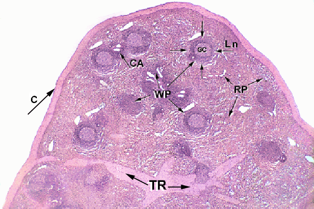
  • Spleen: Can look similar to the other organs (lymph node, liver, etc.), BUT it is the only one that has lymph nodules spread throughout the entire organ (lymph node only has them around the outside, etc.). Remember that the spleen has unique cells in the germinal center (t & b cells). 
Spleen -- see the lobules spread all over? 
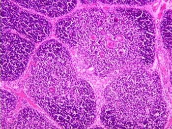
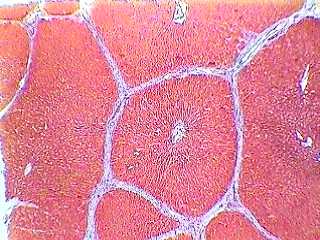
  • Thymus: LOBES that have the characteristic darker outside with lighter inside. Hassall's corpuscles (noticeable cells at the center of the germinal center). Be careful, the thymus can look very similar to the liver, but is distinguishable because the liver does not have the lighter centers and darker outsides of the lobes. The liver can have vasculature running down the center of the lobes which can look like Hassall's corpuscle so keep that in mind for the midterm/final! Use the shading of the lobes as your guide. See how the bottom pictures look similar??? The one on the right is the liver, the left is the thymus. The point of differentiation is the shading of the lobes. 
Hassall's Corpuscle - See how it looks different than vasculature? (no noticeable lumen, no RBCs
Can you tell which is the liver and which is the thymus?
Hint: this one's the liver
  • Tonsils: Associated epithelium is stratified squamous , though not always visible. TONS of nodules, generally arranged in rows. 


Stuff from this section that won't be u-finds:
-Reticulocytes -Eosinophil -Basophil -Monocyte -Megakaryocyte -Lymphatic vessel Stuff from this section that you won't need to know for this week (but will in the future):
N/A
Stuff from this section you don't need to know for lab:
-Pages 28 and 31 are helpful to know but you will not be quizzed on them directly


Terms List: (key: know the histological names for the structures with [h] after them; assume you must be able to find these structures on your own slides unless indicated by [x])

***This list is not guaranteed to be exhaustive, and only includes terms from this week. While we will not directly quiz information from previous weeks, knowledge of previous material will help a lot***


***Make sure you know everything on the chart that was on the board about WBCs***

RBC (erythrocyte)
                Erythropoiesis
Reticulocyte [x]
***A note about the WBC’s: You should be able to find a neutrophil and lymphocyte on your own blood slide. It might be harder to find the other three WBC’s. With that said, your blood smear slide in your box will oftentimes have the others as well.
Neutrophil
Eosinophil
Basophil
Monocyte
Lymphocyte
Megakaryocyte [x]
                Thrombocyte
                Thrombopoiesis
Lymph node [h]
                Medulla
                Cortex
                Lymph nodule [h]
                Germinal center [h]  – what cells are found there?
Ileum
                Peyer’s patches [h]
                Germinal center [h]
Appendix [h]
                Lymph nodule [h]
                Germinal center [h]
Spleen
                White pulp [h]
                Red pulp
                Trabeculae [h]
Thymus 
                Cortex [h]
                Medulla
                Interlobular septa [h]
                Hassall’s corpuscle
Tonsil
                Lymph nodule [h]
                Germinal center
                Epithelium [h]
Lymphatic vessel [h] [x]

Thursday, February 2, 2023

Connective Tissue Stuff

Good news! Connective tissue is definitely a lot less material than the last couple of weeks have been. For this section, approaching the histological naming portion thinking about the idea that "form follows function" will help you. On the quiz for this week, don't worry about writing out the full histo. name (e.g. dense collagenous connective tissue regularly arranged), the abbreviation is totally fine (e.g. DCCTRA). Knowing what the acronym stands for will help you big time in remembering what kind of function it has. Always make sure you know all of the bolded terms in each chapter.

Key things to remember:
-DCCTRA is by far the most common type of connective tissue in the organs that we are studying this week, if you can't remember what kind of connective tissue is present in a specific organ, you're best bet is to go with DCCTRA.
-Make sure you can identify the different fibers in areolar tissue (collagen, elastic) and also be able to ID fibroblasts and know what they secrete. Your manual gives a good explanation of this if you have any questions.
-Hyaline cartilage and elastic cartilage BOTH have chondrocytes, chondroblasts, and a perichondrium (layer on the very outside of the cartilage). The ONLY way to tell them apart is that elastic cartilage has elastic fibers (why it's DECTRA) which are only visible if the cartilage is stained with Orcein-Resorcein/Verheoff's stain. The elastic fibers are very dark because of the stain. Don't get confused though because hyaline cartilage can be stained darker (e.g. purple) too, but it will be a uniform darkness. See below for pics of how elastic fibers cause a non-uniform darkness from the stain.


Examples above are all Elastic cartilage. Notice the contrasting for the elastic fibers and the extracellular matrix.

hyaline cartilage slide
Hyaline cartilage. Notice how the extracellular matrix (space outside of chondrocytes) is much smoother? No elastic fibers.

-Fibrocartilage has NO perichondrium, and most often does not have a noticeable chondroblast layer (though it can...). It will have visible chondrocytes if zoomed out, and a meshwork of irreg. fibers if zoomed in. Look in your lab manual for a good example of both. If we show you fibrocartilage on a slide question, we'll show you the edge of it and it'll look like it just ends, with no perichondrium. 

Fibrocartilage. Notice the lack of a perichondrium/chondroblasts.

-Know where to find lamina propria! In the intestinal tract (just deep to the epithelium layer before you get to the submucosa) and in the oviduct (also just deep to the epithelium layer). 
-The hepatic/glisson's capsule surrounding the liver is DCCTRA as are almost all capsules. Students are often confused when they see a zoomed out picture of the liver. It has many lobules and they are very noticeable when zoomed out. Don't get confused if you see that!


See the lobules? The capsule is labeled C, and B are the interlobular septa. Which are also DCCTRA


-Tendons have a general waviness to them which is especially noticeable when zoomed out further than the picture in the lab manual and they help to connect muscle to bone. They can be other colors than pink as is shown in the manual. They have NO horizontal striations which makes them look different than muscle. 


Both photos above are of tendon. Compare them with the one below of skeletal muscle. See the horizontal striations in the one below? That's one key way to tell them apart.


-Ligaments - Don't stress too much about them. Know that they connect bone to bone and that they have elastic fibers (thus DECTRA instead of DCCTRA). What stain would you use to make the elastic fibers visible?
-Know all the terms in the section about bones....


Before you start naming zones, make sure you always orient yourself - which side is cartilage, which is bone???
(In this one, cartilage is on the left, bone is on the right.)


Again, which side is bone, which is cartilage?
(Cartilage is on the left, bone is on the right where it gets redder. Ignore the black boxes drawn on there.)

-Make sure you can find endochondral bone formation before your quiz! Each box has an endochondral bone slide, but it is often hard at first to find where in the slide the endo. bone formation is occurring. These are easy points as long as you can find it on your slide!
Remember: Real
                   People
                   Help
                   Cancerous
                   Rats
Some hints for each zone: zone of reserve (think reserved people stay by themselves) has mostly single/double cells that are fairly flat. Zone of proliferation (think, they are prolific and having lots of kids...) shows mostly flat cells, but they are arranged in columns with several others. Zone of hypertrophy, the number of cells in the columns don't change, but they are much bigger (more swollen looking). This layer is probably one of the best landmarks because it is usually the most visible. Zone of calcification is immediately after the zone of hypertrophy where there are no more visible cell membranes because the cells are becoming calcified. If asked to identify it, point immediately after the zone of hypertrophy or else you risk pointing to the zone of resorption. If asked to point to the zone of resorption, make sure you are not right next to the zone of hypertrophy.
-For the developing tooth, pick a landmark! Dentin is often the most recognizable and is always thick and colorful (usually bright pink). Or, you could use enamel - it's always the darkest layer. Make sure to know that the ameloblasts secrete enamel inward toward the tooth and that odontoblasts secrete the dentin outward. Know what the ameloblasts and odontoblasts are histologically, and make sure that if you are asked to point them out, that you are pointing to the right layer! They are fairly obvious because they are and look like simple columnar! Sometimes there are spaces in between layers that aren't layers, but they can throw you off. Beware. 
 
All pics of developing teeth. Notice how the dentin layer is the most obvious landmark in each?? (left=the lighter pink, smooth layer; right=bright pink thick layer)  I would suggest identifying the dentin layer and then working out/in from there.

-For the spleen, look for a thick capsule with lymphatic nodules as your landmarks. NOTE: do not think that white pulp is what looks white and the red pulp is what's red! The white pulp is what is inside the lymph nodules (see the pics below) and the red pulp is everything surrounding the lymph nodules. Not confusing at all.
See the thick capsule? The circular parts are the lymph nodules.

Where's the red pulp? White pulp?
(In this one, the red pulp actually does look pretty red)

-The internal elastic laminae is the squiggly dark line (stained with orcein-resorcein) you see near the lumen of arteries (most obvious on medium sized arteries) and the external elastic laminae is the collection of dark squiggly lines a bit deeper once you get past the smooth muscle. The tunica adventitia is immediately after the external elastic laminae. When the artery is in diastole (relaxed), the squiggly lines are also relaxed and they remain squiggly -- DECTIRA (squiggly lines are irregularly arranged...). When the artery is in systole (blood is higher pressure), the elastic laminae are stretched and are no longer squiggly -- DECTRA (regularly arranged). It's helpful to remember that YOU WILL NEVER see a slide of an internal or external elastic lamina in systole (the animals are no longer living, thus their hearts are no longer pumping).


What is the internal elastic lamina histologically both in diastole and systole?


Can you identify the internal & external EL's as well as the Tunica Adventitia? What are their histological names?
-Finally, make sure you can find some examples of adipose (unilocular adipocytes) on your own slides. Some good places to check are on your parotid slide or around vasculature.

Good luck this week! Let us know if you have any questions.


Stuff from this section that won't be u-finds:

-Reticular fibers -Elastic cartilage -Fibrocartilage Stuff from this section that you won't need to know for this week (but will in the future):
-Muscularis mucosa

Stuff from this section you don't need to know for lab:
-The whole page 22 is helpful to know, but you won't be quizzed on in directly -Ligament (we won't test you on it)


Connective Tissue Key Terms
Key: Know the anatomical and histological names (including modifications) for the following bolded structures; assume that you will be required to find the structures indicated by * on your own slides.
***This list is not guaranteed to be exhaustive, and only includes terms from this unit. While we will not focus on quiz information from previous weeks, knowledge of previous material may be useful***
Focus on connective tissue histology, but keep in mind all of the following terms.
Basic Connective Tissues
Aerolar Connective Tissue
·       Aeolar connective tissue*
·       Fibroblasts*
·       Collagen fibers*
·       Elastic fibers*
Hyaline Cartilage
·       Hyaline cartilage*
·       Chondroblast layer*
·       Chondrocytes*
·       Nest cells*
·       Perichondrium*
Elastic Cartilage
·       Elastic cartilage*
·       Chondrocytes*
·       Chondroblast layer*
·       Perichondrium*
Fibrocartilage
·       Fibrocartilage*
·       Nest cells*
·       Chondrocytes*
Digestive System
Intestinal Tract
·       Lamina propria*
·       Muscularis mucosa
·       Submucosa*
Liver
·       Glisson’s capsule*


Musculoskeletal System
Tendon
·       Tendon*
·       Collagen fibers*
·       Fibroblast nuclei*
Ligament
·       Ligament
·       Elastic fibers
·       Fibroblast nuclei
Bone
·       Haversion canal*
·       Osteocytes in lacunae*
·       Canaliculi*
·       Concentric lamellae*
·       Interstitial lamellae*
Endochondral Bone Formation
·       Zone of reserve*
·       Zone of proliferation*
·       Zone of hypertrophy*
·       Zone of calcification*
·       Zone of resorption*
·       Perichondrium*
·       Periostium*
Developing Tooth
·       Dental pulp*
·       Odontoblast layer*
·       Predentin*
·       Dentin*
·       Enamel*
·       Ameloblast layer*
Respiratory System
Trachea
·       Chondroblast layer*
·       Perichondrium*
·       Tracheal epithelium
Dermis
·       Dermis*
·       Epidermal epithelium


Lymphatic Tissue
Spleen
·       Splenic capsule*
·       White pulp*
·       Red pulp*
Lymph Node
·       Reticular fibers
·       Dense lymphatic tissue
Cardiovascular
Blood Vessels
·       Internal elastic lamina (systole and diastole)*
·       External elastic lamina (systole and diastole)*
·       Tunica adventitia*
Miscellaneous
Adipose Tissue
·       White adipose cells*


Oviduct
·       Oviduct epithelium
.        Lamina propria*